Cataract – diagnosis
The cataract is an eye disease that develops slowly over many years. At the beginning of this widespread eye complaint it is particularly easy to overlook initial signs pointing to the cataract. In older people in particular, the symptoms are often misinterpreted and mistakenly attributed to a general deterioration of health. People who still work, quite often attribute their symptoms to exhaustion or excessive stress. But a cataract must not be taken lightly. In serious cases, if left untreated, it can lead to blindness. Regular medical check-ups with an eye specialist are therefore very important. If diagnosed early, a straightforward operation can help avoid restrictions to everyday activities and a loss of quality of life.
How does a cataract manifest itself?
There are individual differences. Not every patient has the same symptoms, and the course of the disease can vary. As the disease progresses, the cataract increasingly impairs your eyesight. The typical clouding of the lens which is visible to everyone happens at a very advanced stage.
The first signs of a cataract can be quite nonspecific and are not always associated with the disease. Many sufferers are sensitive to bright light and are easily dazzled, such as by bright sunshine, by oncoming traffic in the dark or by flashlight. It also takes longer for the eye to adjust to new light conditions. Even during the early stage, sufferers have problems when switching from bright to dark rooms and vice versa. Some patients experience a temporary reversal of their vision. People who used to be long-sighted suddenly become short-sighted but can see things better nearby. Depending on the type of cataract, people may experience double vision.
Often, contrasts are no longer clearly perceived, outlines become blurred and so-called halos form around sources of light. The patient’s general vision deteriorates – reading and watching TV becomes increasingly tiring. Many people begin to feel insecure in traffic. As the disease progresses, sufferers see everything through a foggy veil that becomes increasingly denser. This is the stage in which colour perception also changes. Colours become paler and lose their luminosity. These visual changes go hand in hand with a deterioration of spatial perception, which also significantly impairs orientation. This can go as far as sufferers reaching for something and missing it. Finally, in the end stage the world disappears in a dense fog and only shadowy outlines are perceived.
How does the eye specialist diagnose a cataract?
Initially, the cataract does not always exhibit clear symptoms, which is why only an examination by an ophthalmologist can provide certainty. In our practice, we use a wide range of different powerful procedures to diagnose the disease, from the slit lamp examination and biometrics to optical coherence tomography (OCT) and all the way to state-of-the-art high-tech diagnostic methods such as 3D cataract analysis using Scheimpflug imaging. An early diagnosis is essential for optimal treatment planning and tells us about the right time for surgery. At the beginning of the disease, there are few procedures that unambiguously diagnose the cataract.
3D cataract analysis / Scheimpflug imaging
When it comes to the early detection of serious eye diseases such as cataracts or pathological changes to the cornea (keratoconus), Scheimpflug imaging is a modern examination technique in eye diagnosis. It maps the entire anterior segment of the eye in three dimensions, making it easier to examine it. We use it to precisely analyse both the anterior eye chamber as well as cornea, iris and lens. The procedure enables an exact quantification of the cataract and thus reliably detects possible corneal clouding. The high-resolution digital images can be rotated and viewed from all sides. This gives the doctor unique insights, and even the smallest changes to the lens do not remain unnoticed. This allows us to detect cataracts early and therefore provide the best possible treatment. The measured values are stored and allow us to monitor tiny changes to the modified eye segment and the lens. This in turn allows us to precisely quantify and classify the gradual changes. This computer-aided process is also ideal for post-surgery check-ups. It tells us about the correct position of the implanted lens and allows us to monitor the healing process.
Scheimpflug imaging is a contact-free examination, and it does not affect the patient in any way. The measurement is performed using a computer-aided high-tech camera, the Pentacam HR AXL Premium. This latest-generation diagnostic tool with its automatically rotating camera performs measurements of the anterior segment of the eye in seconds. The rotation principle with its great number of measuring points provides an extremely high-resolution image of the central cornea. The three-dimensional measurement also allows for a topographical representation of the anterior and posterior corneal surface. This gives us more measurement data and thus a more accurate result.
Optical coherence tomography (OCT)
This modern measuring technique works with laser light and is able to represent different structures of the eye in high-resolution cross-sections and analyse them. In a sense, it gives us insight into the depth of the tissue. Prof. Marcus Kernt’s practice has an anterior segment OCT (Visante OCT with Atlas Topographer, Zeiss Meditech) for the planning of refractive and lens surgery, which is used to three-dimensionally analyse the fine structure of the cornea, the anterior eye chamber, iris and lens. The practice also has a high resolution spectral domain OCT for the detailed examination of the ocular fundus, including the retina and the optic nerves.
Optical biometry
Before a cataract operation can be performed, it is necessary to determine the refractive power of the artificial lens (intraocular lens) that will replace the natural eye lens. This way we can find the perfect lens implant for the patient’s needs. Optical biometry is currently the most precise and most state-of-the-art procedure for this purpose. In contrast to the standard ultrasound method, optical biometry performs the measurement without touching the eye, which means there is no need for a local anaesthetic of the eyeball. The contact-free examination means that the procedure is risk-free, painless and poses no adverse effects.
Optical biometry uses state-of-the-art computer-aided laser technology to make the required calculations. The measurements are carried out in a single step and include all the data, which later results in the best possible visual performance. They include axis length, corneal radius, corneal diameter and, if necessary, the depth of the eye’s anterior chamber. Although the well-established standard ultrasound method paid for by the health insurance companies brings good results, optical biometry is far more accurate. In our practices, we use the IOL Master by Zeiss and the Pentacam AXL.
Contact
We are happy to advise you!
Prof. Dr. Marcus Kernt
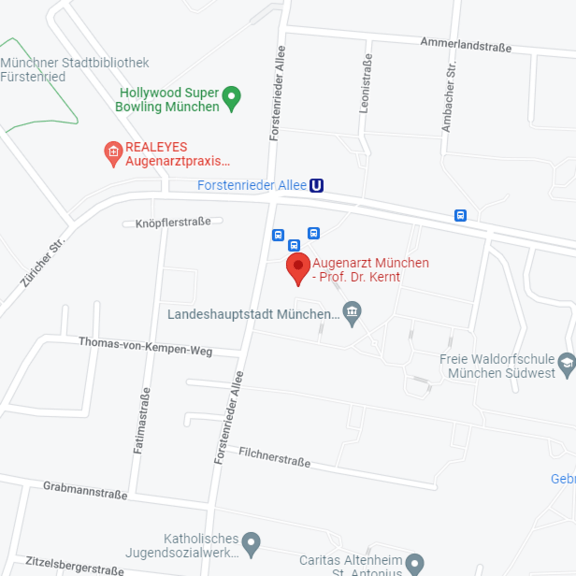
Forstenrieder Allee 59
81476 München


